Welcome To Our Blog!
Welcome to the Cary Eye Center Blog. stay Tuned for new posts and the latest news!
The normal retina is shown in Fig. 1. The round optic disc is seen with normal appearing blood vessels coming out and a normal retina and macula (the center part of the retina that corresponds to our clearest, central vision). The retinal veins are darker and larger than the retinal arteries.
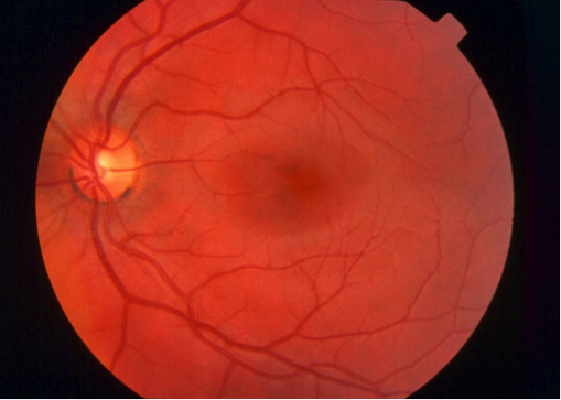
Figure 1
In Fig. 2, we can see some of the early signs of Diabetic Retinopathy. The scattered red spots are called dot and blot hemorrhages and there are some tiny spots that are called microaneurysms. The blood vessels also begin to become abnormal, in this case the white rectangle is showing “venous beading”. Also, some of the small white spots represents exudates and the larger white areas correlated to damage of the nerve fiber layer, called cotton wool spots. Each of these findings correlates to the first stage of Diabetic Retinopathy, called Non-Proliferative Diabetic Retinopathy, or NPDR.
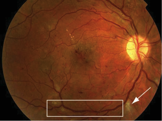
Figure 2
As diabetic retinopathy progresses, it changes from Non-Proliferative to Proliferative Diabetic Retinopathy, or PDR. In Fig. 3, we can see the changes associated with this condition. New, abnormal blood vessels actually begin to form, a process called neovascularization (NV). These can occur near the optic disc (NVD) or in other parts of the retina (elsewhere), termed NVE. In Fig. 4A, some of these acronyms are noted in the diagram. PDR is a very serious condition and can cause hemorrhaging, retinal swelling, scar tissue formation, and retinal detachment. In addition to the aforementioned blood sugar control, laser treatment must be initiated for PDR, as well as sometimes for severe NPDR. The laser treatment is applied to the peripheral retina in little spots, as shown in Fig. 4B. This treatment, while not a cure, can help reduce the ischemic load of PDR and slow down its progression. It will also help to prevent another complication of PDR which is called neovascular glaucoma.
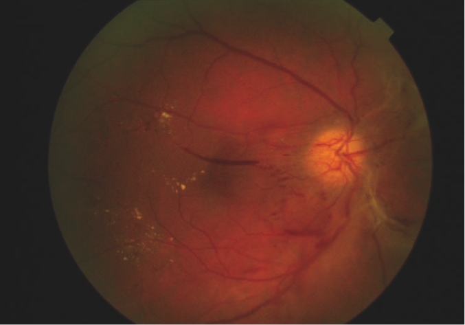
Figure 3
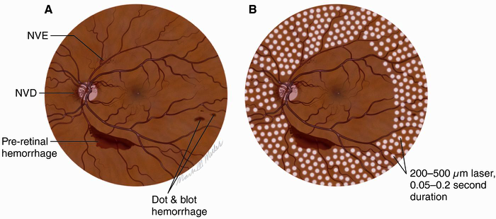
Figure 4
PDR often results is severe hemorrhage that can move out of the retina and into the center part of the eye, called the vitreous. When this happens, it is termed a Vitreous Hemorrhage (VH) (Fig. 5). The dark red spots are the VH. If severe enough, the VH can fill the entire eye, making the person effectively blind. This blood can usually, however, be removed with a surgical procedure.
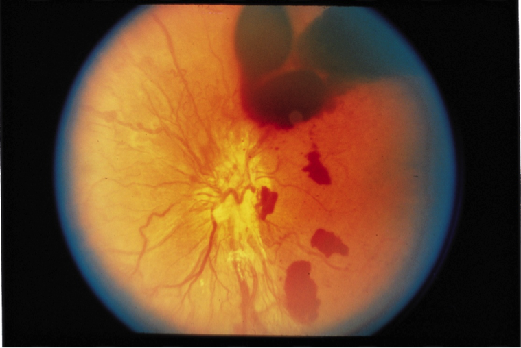
Figure 5
A final visual problem associated with Diabetic Retinopathy is swelling in the center part of the retina, the macula, where our central vision is located. This is termed Diabetic Macular Edema (DME). The retina is normally flat, akin to a dry sponge. Due to leakage of fluid, diabetics can experience swelling in the macula, which causes blurry vision. A diagram of this condition is shown in Fig. 6. The top part of this figure shows a diagram of the anatomy of the macular in DME (Fig. 6A). Below it, there is a real OCT image of a swollen macula with DME (Fig. 6B). OCT, which stands for Ocular Coherence Tomography, is a relatively new technology that takes a “slice” of the retina to determine the health of this detailed anatomy. The large black cystic spaces are representative of fluid accumulation, like a wet sponge. Below this image is an OCT of a normal macula (Fig. 7) for comparison.

Figure 6
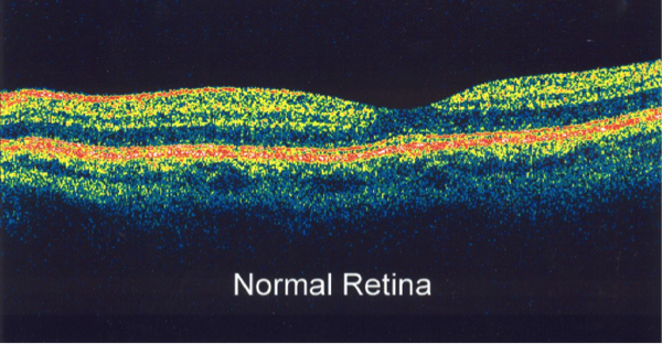
Figure 7
Figures 1-7 are © 2015, American Academy of Ophthalmology.
Yesterday, I had a frightening eye situation with my wife. Cary Eye Center took my wife without an appointment and the whole staff was wonderful. The icing on the cake was Dr. O’Neal. I examined his training history prior to calling the office, and I was very impressed. Meeting Dr. O’Neal and experiencing his reassuring, extremely professional demeanor was just what my wife needed, to allay her anxiety. This visit convinced us that all of our future eye treatment will be done by Dr. O’Neal. In short, we cannot recommend an ophthalmologist higher!

I recently had occasion to meet Dr. O’Neal, under rather inauspicious circumstances. While performing a home improvement project, I sustained a fairly serious injury to my left eye, requiring a visit to the emergency room. Fortunately for me and for the future of that eye, Dr. O’Neal was on call that day. His swift care, attention, and great competence resulted in the best possible outcome in an otherwise doubtful situation. Moreover, his obvious concern for my well-being and clear explanation of the circumstances attendant to the injury (I, being a layman) allowed a scary situation to feel less so. He has a new patient in me and my family, and I couldn’t more enthusiastically recommend him to anyone in search of a great eye-care provider.
“I have been a patient of Dr. O’Neal’s for over a decade. When I first started seeing him, my eyes were a mess… Working with Dr. O’Neal, he was able to systematically diagnose the issues and treat them. My eyes have never felt better! At one point in my treatment, I ended up having an allergic reaction to one of the eye drops. It was an uncomfortable night to say the least. I called his office when it opened and he was able to fit me in that morning and fixed the problem. It was very much appreciated! The new office is really nice and the staff have been very friendly. Overall, my eyes have never felt better…and I’m very happy to be Dr. O’Neal’s patient.”
“Dr. Kevin O’Neal restored my faith, and I will always remain grateful to him…His service answered and said they would relay the message to him. To my amazement, within minutes, as I was on my way out of the door, my phone rang and it was Dr. O’Neal himself, personally returning my call at 10PM in the evening. By now I had completely lost vision out of my right eye and thought I was going to be permanently blind in that eye. Dr O’Neal comforted me on the phone with his words and told me he would be waiting for me to arrive at the hospital. He was there when I arrived and I was given IMMEDIATE treatment because of him. I did not have to wait my turn. He performed emergency surgery that evening and restored my vision and saved my eye.”
“I have been a patient of Dr. O’Neal for 10 years. I have always received, in my opinion, the highest qualityof medical care. His medical skills and competence is matched by his professionalism. Dr. O’Neal embodies a friendly personality and compassionate attitude, which immediately puts patients at ease. I have recommended Dr. O’Neal to many friends and will continue to do so.”
100 Parkway Office Court, Suite 200
Cary, NC 27518
Mon-Thu: 8:30a-5:30p (closed 12:30-1:30)
Fri: 8:30a-1:30p
Sat & Sun: CLOSED
Welcome to the Cary Eye Center Blog. stay Tuned for new posts and the latest news!